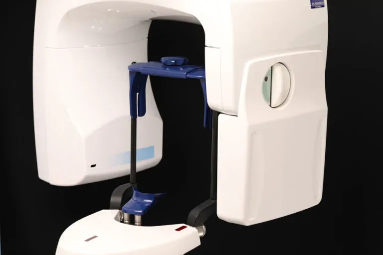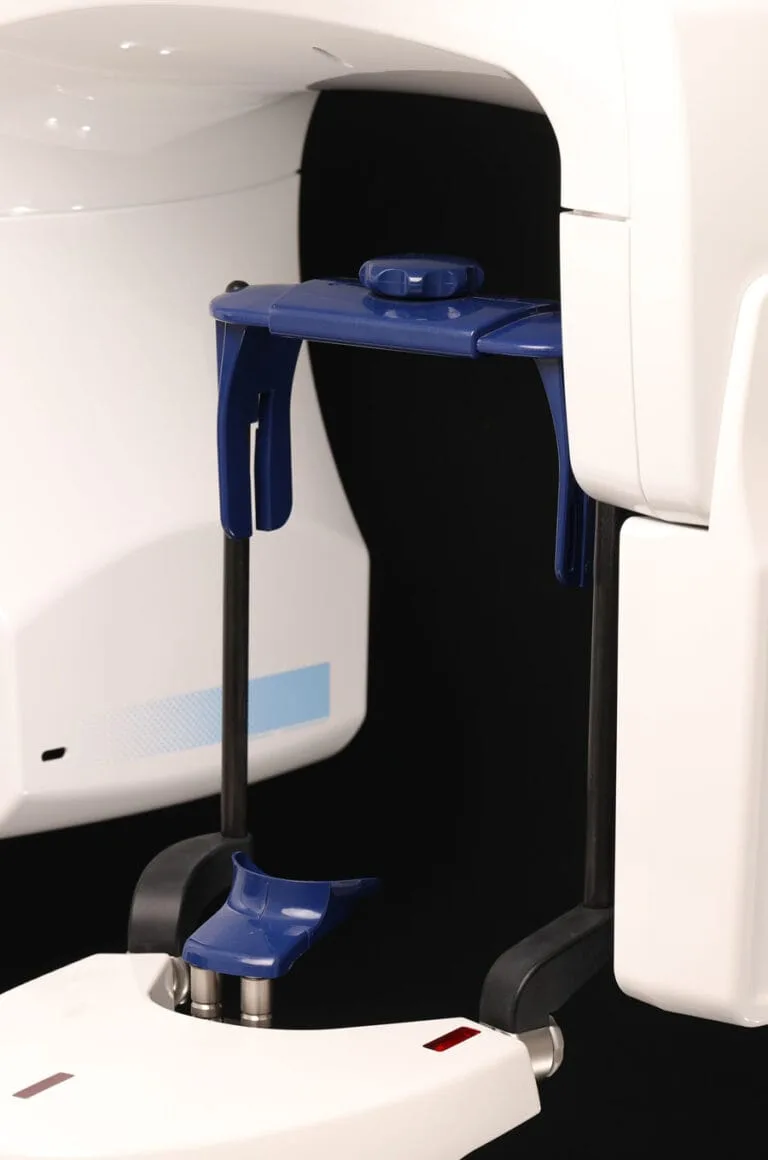
A dental X-ray is the basic diagnostic examination that lets me see what the naked eye and a probe cannot: decay between the teeth, inflammation at the root tip, the state of the bone around a tooth or an implant. In my practice I use digital sensors, so the image is on the screen within seconds and the radiation dose is small. This page mainly describes the small intraoral images of individual teeth; the overview of the whole jaw on one image is described on the panoramic X-ray page, and three-dimensional imaging on the 3D dental imaging page.
Who it is for
An intraoral X-ray makes sense when:
- one particular tooth hurts or is sensitive and I want to see the root and the bone around it (periapical image),
- we are looking for decay between the teeth that cannot be seen during the examination (bitewing image),
- we are checking whether a filling or crown is leaking under its margin,
- during root canal treatment we measure the length of the canals and check the filling,
- we monitor the bone around an already placed implant at the annual check-ups,
- in gum disease we assess how much bone has been lost around an individual tooth.
When an intraoral image is not the right choice:
- if we need an overview of all the teeth, the wisdom teeth, the jaw joints or the sinuses, the correct examination is a panoramic X-ray,
- for implant planning an intraoral image is not enough, because it does not show the width of the bone; there a 3D CBCT scan is needed,
- during pregnancy we only take X-rays when they are necessary for treatment, and always with a protective apron; non-urgent images are postponed until after the birth. If you are or could be pregnant, please tell me before the image is taken.
How it goes
- Conversation and clinical examination (5–10 minutes). I only take an image when it changes the decision about treatment; I do not take routine images “just in case”.
- Preparation: you remove glasses, jewellery and removable dentures and put on a protective apron.
- The image: I place a small digital sensor in your mouth next to the tooth being imaged and you hold it for a moment with your bite or a finger. The exposure lasts less than half a second, the whole image less than a minute. For a bitewing you bite on a plastic holder.
- Looking at the image together: the image is on the screen immediately, I show you what I see and explain what it means for your treatment.
- Plan: the image becomes part of your record so that we can compare changes at your next visits. On request you receive it in digital form for another doctor or your insurer.
Altogether, including the examination, it usually takes 15–20 minutes and is done at the same visit as the treatment.
Price
| Service | Price | What it includes |
|---|---|---|
| Dental X-ray (intraoral or panoramic image) | 25–80 € | digital image, reading and explanation, image in your record |
| Panoramic X-ray (OPG) | 25–80 € | overview of the whole jaw on one image |
| 3D CBCT | 80 € | three-dimensional scan for implant planning |
| Filling | 80–130 € / tooth | the most common treatment after decay is found |
A small intraoral image is at the lower end of the range, a panoramic image at the upper end. If you come for a first visit, the image is already included in the price of the visit: 50 € with a panoramic X-ray or 80 € with a 3D CBCT; at the first visit I also prepare a written treatment plan with the final price, which is not binding for you. Part of your dental costs can be claimed in Slovenia as a special tax relief; check the conditions with FURS, as the rules change.
Afterwards
There is no recovery and no restriction after a dental X-ray; you can eat, drive and go back to work. The dose from one intraoral image is roughly what you receive from the natural environment in a few hours, so there is nothing to “make up for” and no reason to avoid it at your next treatment.
How often to take images: with healthy teeth and no complaints, bitewing images roughly every 2–3 years are enough; with a high risk of decay or after extensive work, more often. With implants I take a control image when the crown is fitted and then at the annual check-up, so that any bone loss is seen in time. I keep the images, so that when you change dentist the X-rays are not repeated unnecessarily.
Why at Koderman
- Digital sensors and a panoramic unit in the same practice: the image is taken during the examination, not by referral two weeks later.
- We look at every image together on the screen; you decide about treatment with understanding, not on trust.
- When the image shows that an implant is needed, the next step, a 3D CBCT scan, is available in the same building.
- Appointment usually within 7–14 days, no waiting list.
- Examination and explanation in Slovenian, German, English and Croatian.
Frequently asked questions
How much does a dental X-ray cost?
From 25 to 80 €, depending on the type of image: a small intraoral image is cheaper, a panoramic image more expensive. At the first visit the image is already included in the price of the visit (50 € with a panoramic X-ray, 80 € with a 3D CBCT).
Is a dental X-ray dangerous?
The dose from a digital intraoral image is very small, comparable to a few hours of natural background radiation. Even so, we do not expose ourselves to radiation without reason: I only take an image when it affects treatment, and we use a protective apron.
Can I have a dental X-ray during pregnancy?
During pregnancy we postpone non-urgent images. If an X-ray is needed because of pain or infection, we take it with protection; the dose from a tooth image is very far from the abdomen and very small. Always tell me about a pregnancy in advance.
What is the difference between a dental X-ray, a panoramic X-ray and 3D imaging?
An intraoral image shows one to three teeth very precisely. A panoramic X-ray shows all the teeth, both jaws and the joints on one two-dimensional image. A 3D CBCT scan shows the bone in all three dimensions and is the only one that is sufficient for safe implant planning.
Does it hurt?
No. The only discomfort is the sensor in the mouth, which triggers a gag reflex in some people; when imaging the back teeth we reduce it with correct positioning and quick work.
Can I bring an X-ray from elsewhere?
Yes. If you have a digital image up to about six months old, I will use it and will not repeat the X-ray unless changes make it necessary.